
Medical X-ray imaging in Rockaway, NJ (or elsewhere more relevant) produces a picture that helps doctors find broken bones, tumors and other abnormalities. It only exposes your body to radiation for a very short time. X-rays pass through the body onto specially treated film or digital media, producing a black-and-white image of internal structures. The more dense a form is, the whiter it appears on an X-ray.
C-arm
Doctors can see hard and soft tissues in your body during this imaging test. Dense materials like bones absorb X-rays and appear white on the image, while soft tissue allows X-rays to pass through and appear black. It helps your doctor to diagnose health problems.
Before an X-ray exam, you should remove any jewelry or body piercings and may be asked to wear a gown. X-rays carry a small risk, but the benefits outweigh the risks. If you’re using a mobile C-arm for fluoroscopy, consider a system with an adjustable source-to-image distance from https://www.minicarm.com/.
It will help reduce scatter radiation exposure for staff members. A medical physicist can advise you on this and other safety issues. They can also help you ensure your c-arm setup is safe and compliant with all regulations.
O-arm
The o-arm is an image-guided surgical navigation system that allows surgeons to be more precise. They can fix problems with fewer or smaller incisions and faster patient recovery time. This mobile X-ray system provides high-quality, uniplanar images that are free of distortion and can be recalled at the touch of a button at any time during surgery.
It eliminates time-consuming repositioning and additional X-ray exposure for scout films. With the o-arm, the surgeon can receive a highly detailed 3-D image of the surgical site within seconds, which is ideal for verifying the position and placement of hardware.
It helps ensure that screws are placed correctly and may reduce the need for future revision surgery. Moreover, this imaging system also enables minimally invasive surgery and reduces patient radiation doses.
Periapical
X-rays capture images of the inside of your body. They show bone and tissue and can help your doctor diagnose injuries and illnesses. A periapical X-ray scans one tooth from the crown (chewing surface) to the tip of the root. This X-ray helps dentists see dental caries, periodontal disease, and other teeth, bone and surrounding tissue abnormalities.
Your provider may administer a liquid contrast medium before some types of X-rays. This dye, usually barium or iodine, highlights specific areas on the X-ray image and helps your provider discover more about your condition. The contrast medium can be swallowed or given through an injection or inserted into the rectum (an enema). You may feel flushed or experience a metallic taste after the procedure.
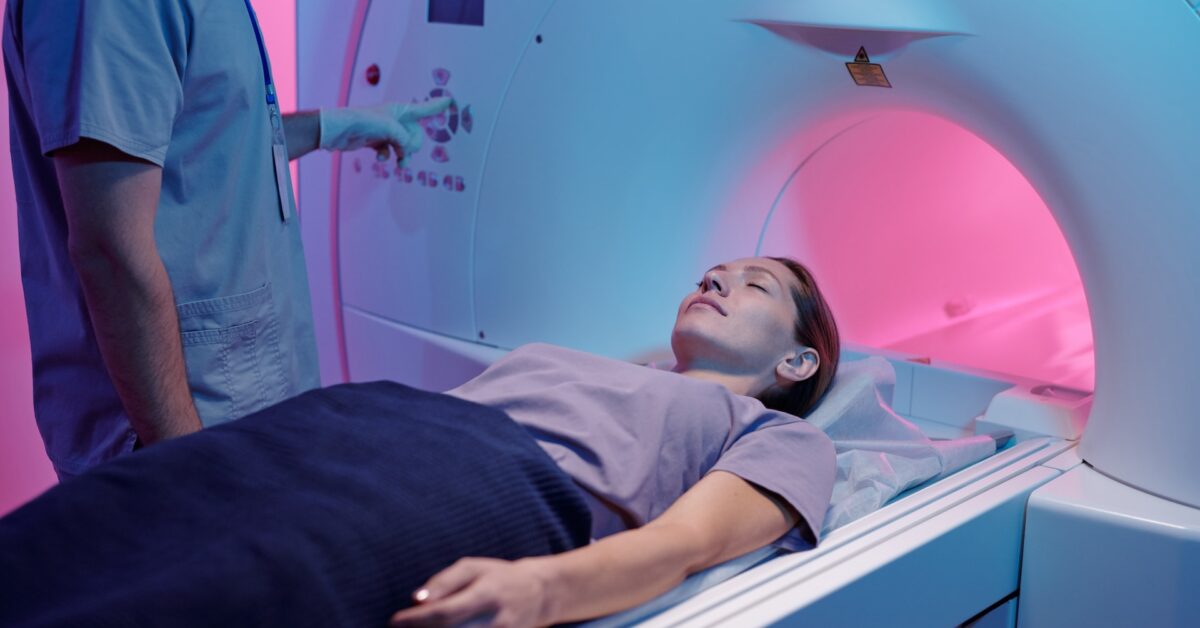
Bone Density Scan (DXA)
X-rays are a useful diagnostic tool for many conditions, from broken bones to life-threatening illnesses. They expose you to a small amount of radiation, but the benefit is worth it.
The bone density test, also known as bone mineral densitometry (DXA), measures the strength and thickness of your bones with low-dose X-rays. It is especially helpful in detecting osteoporosis and measuring the effectiveness of treatment for osteoporosis.
During the DXA scan, you lie on a padded table while an arm passes over your body. It is smaller than a central device and only takes a few minutes. You may be able to keep your clothes on. It is important to let your doctor know if you are pregnant or think you might be pregnant because the amount of radiation used in the test is very small and can pose a risk for pregnancy.
Bitewings
Bitewings are the traditional film of choice for detecting tooth decay between the back teeth and assessing periodontal bone loss. They are especially useful in identifying interproximal caries lesions and the proximal dentin and enamel interface.
During a bitewing radiograph, the patient bites down on a cardboard or plastic tab to hold the mouth together, and a film is exposed by an x-ray tube for a few seconds. The dental professional next examines the generated X-ray to look for indications of tooth decay and other oral health issues, including immature or impacted teeth.
When used judiciously, bitewings are an effective diagnostic tool in detecting caries and are often recommended at repeat intervals. By adhering to ALARA principles, using a high-quality holder and beam-aiming device, practicing precise technique, and avoiding retakes, bitewing radiographs are of optimum diagnostic accuracy with minimal radiation exposure.